


Study of coated magnetosome minerals
1. Manufacture of coated magnetosome minerals
To avoid the toxicity problems related to the presence of endotoxins and immunogenic organic substances at the surface of magnetosome minerals, Nanobacterie researchers have developed a four-stage synthesis method:
- First, magnetotactic bacteria are cultivated in a fermenter (step 1);
- Magnetosomes are next extracted from magnetotactic bacteria (step 2);
- Magnetosomes are purified to remove the residual organic material, which surrounds the mineral part of the magnetosome and consists of endotoxins (step 3);
- Magnetosome minerals obtained after step 3 are then coated with various substances, such as citric acid, poly-L-lysine, polyethylenimine (PEI), carboxy-methyl-dextran, chitosan, neridronate, oleic acid, etc. (step 4).
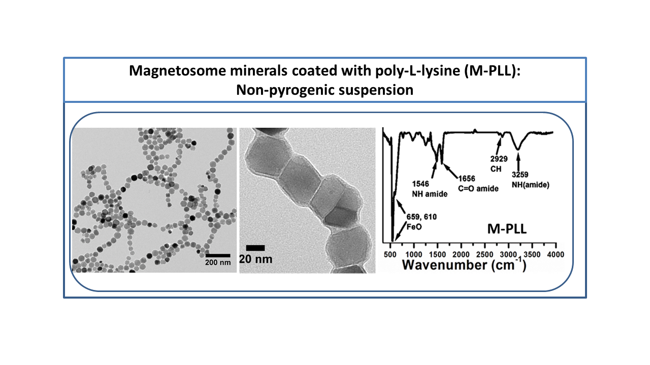
We have shown that coated magnetosome minerals obtained at the end of step 4 are non-pyrogenic, biocompatible, stable in aqueous suspension, and have better heating properties than chemical nanoparticles commonly used for magnetic hyperthermia treatments.
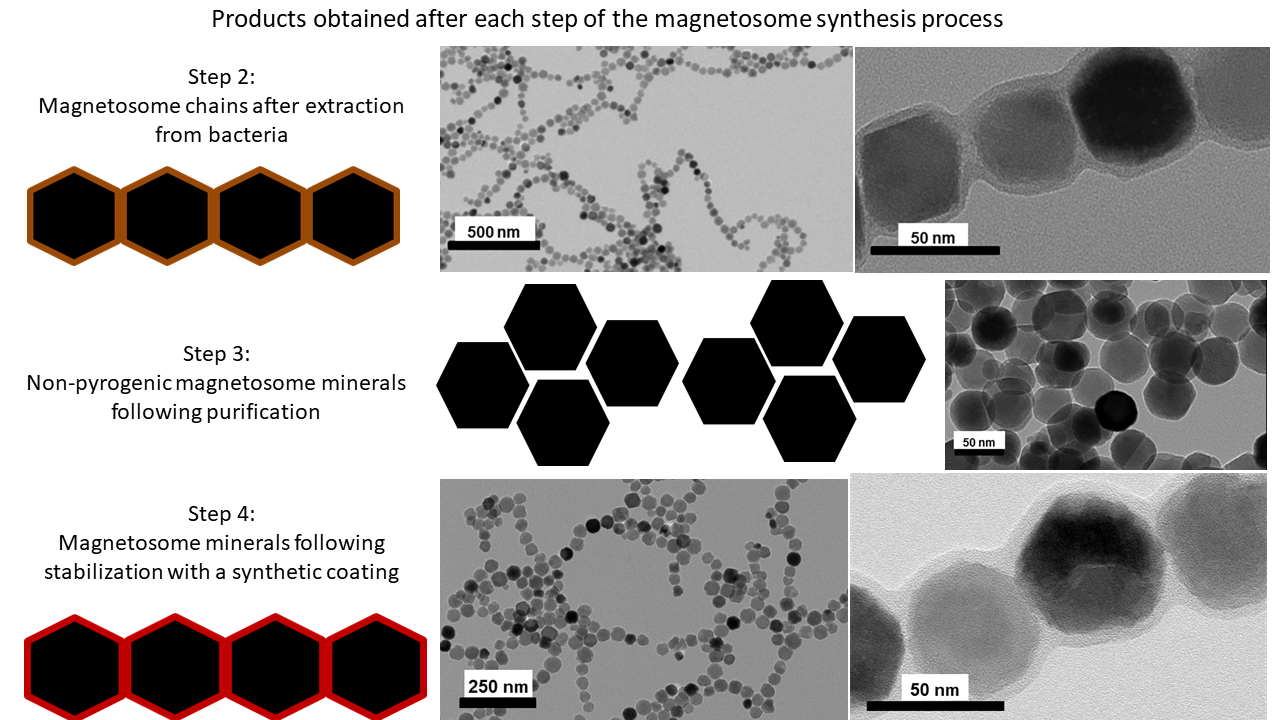
The figure below shows transmission electron microscopy images of magnetosomes obtained at the end of the different stages of treatment:
- Chain of magnetosomes with a natural coating originating from a magnetotactic bacterium (step 2);
- Magnetosome minerals without coating (step 3);
- Magnetosome minerals covered with a synthetic coating (step 4).
2. Organization of coated magnetosome minerals
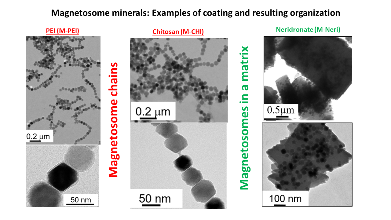
The figure below gives examples of organizations of coated magnetosome minerals obtained at the end of step 4:
- A chain arrangement for PEI and chitosan coatings;
- Encapsulation in a matrix for the neridronate coating.
It is interesting to note that, for some coatings, we observed that the chain arrangement was quite similar to that observed inside the magnetotactic bacteria.
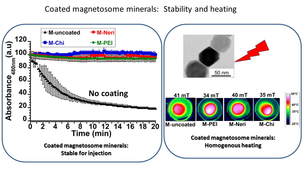
3. Stability and heating properties of coated magnetosome minerals
In addition, the figure below indicates that these coated magnetosome minerals are sufficiently stable (>20 minutes) for injection, and that they allow homogeneous heating, meaning they have favorable properties for magnetic hyperthermia.
4. Toxicity of coated magnetosome minerals
Researchers at Nanobacterie evaluated:
- Magnetosome minerals coated with seven different coating agents all appear non-cytotoxic according to ISO 10993-5, since in each case the percentage of cell inhibition remained below 30%;
- Acute toxicity. A quantity of 8 mg poly-L-lysine-coated magnetosome minerals administered to the tail of mice did not induce weight loss or death, indicating the low acute toxicity of these magnetosomes;
- Pyrogenicity-Limulus amebocyte lysate (LAL) tests reveal comparable endotoxin concentrations for coated magnetosome minerals and chemical nanoparticles.
In addition, pyrogenicity tests carried out on rabbits revealed that poly-L-lysine-coated magnetosome minerals were pyrogen-free.
5. Efficacy of magnetosome minerals coated in subcutaneous GL-261 murine glioblastomas
Nanobacterie researchers have also tested the efficacy of poly-L-lysine-coated magnetosome minerals.

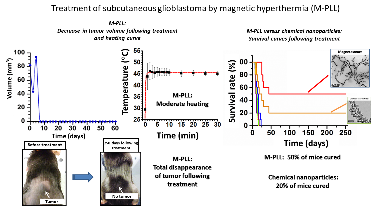
For this purpose, we first implanted glioblastoma GL-261 tumors subcutaneously and, when glioblastomas reached 50–150 mm3, administered approximately 2.5 mg of the magnetosome minerals to the center of these tumors. The tumors were then exposed to 15 applications of an alternating magnetic field of various strengths between 11 and 31 mT and a frequency of 198 kHz to reach heating temperatures of 43–45°C. The treatment led to tumor growth delay in half of the treated mice and to total tumor destruction in the other half. Under equivalent treatment conditions, signs of antitumor activity were much less pronounced with chemical nanoparticles, which yielded total tumor destruction in only 20% of the mice.
The figure below (on the left) summarizes the conditions of treatment with poly-L-lysine-coated magnetosome minerals. The other figure (on the right) compares the survival rates obtained with coated magnetosome minerals and chemical nanoparticles.
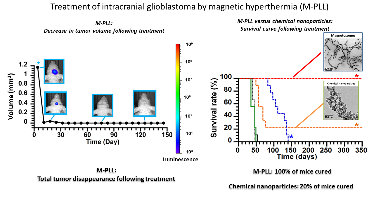
6. Efficacy of magnetosome minerals coated in intracranial U87-Luc murine glioblastoma
Nanobacterie researchers then grew U87-luc glioblastomas within mouse brains. When glioblastomas reached a volume of 1.5 mm3, we administered 0.5–0.7 mg of poly-L-lysine-coated magnetosome minerals to the center of these tumors, and the tumors were exposed to 27 applications of an alternating magnetic field of 27-mT strength and a frequency of 198 kHz. The treatment led to complete destruction of glioblastomas for 100% of treated mice. When carried out under equivalent conditions but using chemical nanoparticles instead of magnetosomes, the treatment was much less efficient, yielding complete tumor destruction in only 20% of the mice.
The figure below (on the left) summarizes results following treatment with poly-L-lysine-coated magnetosome minerals. The other figure (on the right) compares the survival rates obtained with coated magnetosome minerals and chemical nanoparticles.