


Probe based on fluorescent magnetosomes
1. Manufacture of the fluorescent probe
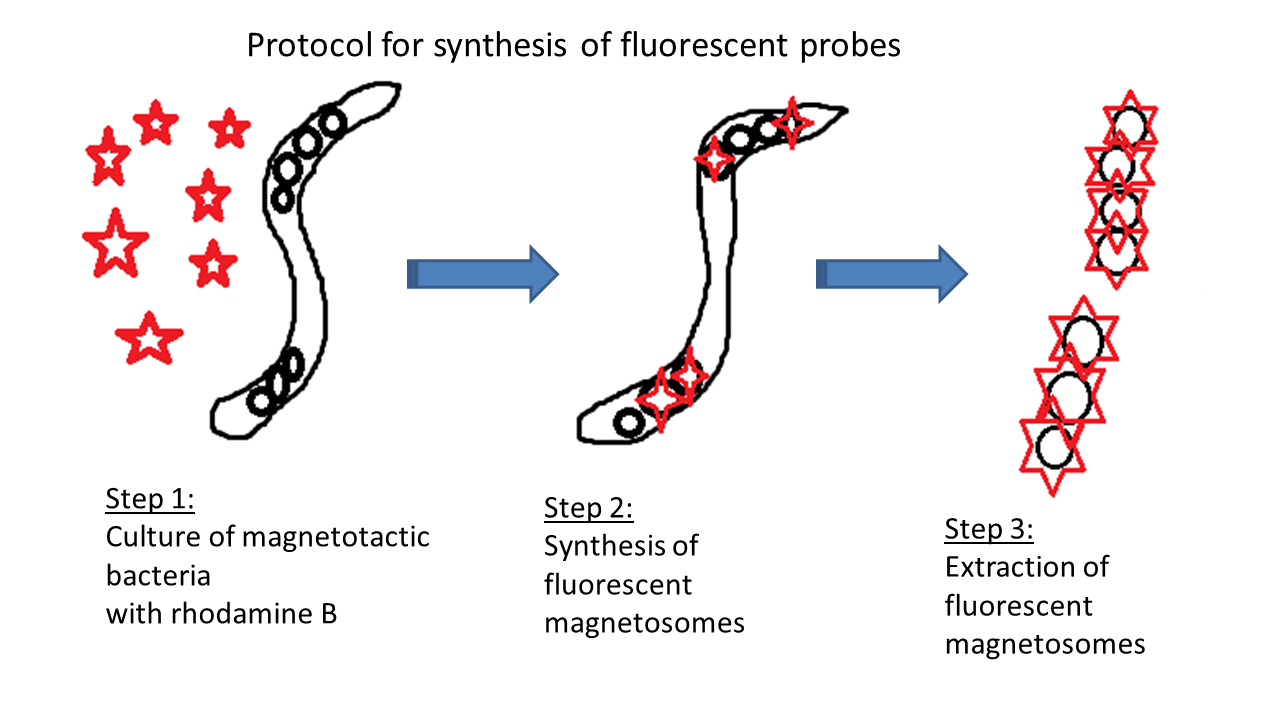
Nanobacterie has developed a method for producing fluorescent magnetosomes that can be used as fluorescent probes. To do this, 400 μM of rhodamine B is introduced to the culture medium of magnetotactic bacteria. Because of its chelating properties, rhodamine B can become incorporated with iron in magnetosomes to form magnetosomes linked with rhodamine B. These magnetosomes are then extracted from the magnetotactic bacteria for use.
The figure below summarizes the main manufacturing steps of this fluorescent probe, which are:
- Culture of magnetotactic bacteria in the presence of rhodamine B (step 1),
- Synthesis of fluorescent magnetosomes by magnetotactic bacteria (step 2),
- Extraction of fluorescent magnetosomes from magnetotactic bacteria (step 3).
2. Operating mechanism of the fluorescent probes
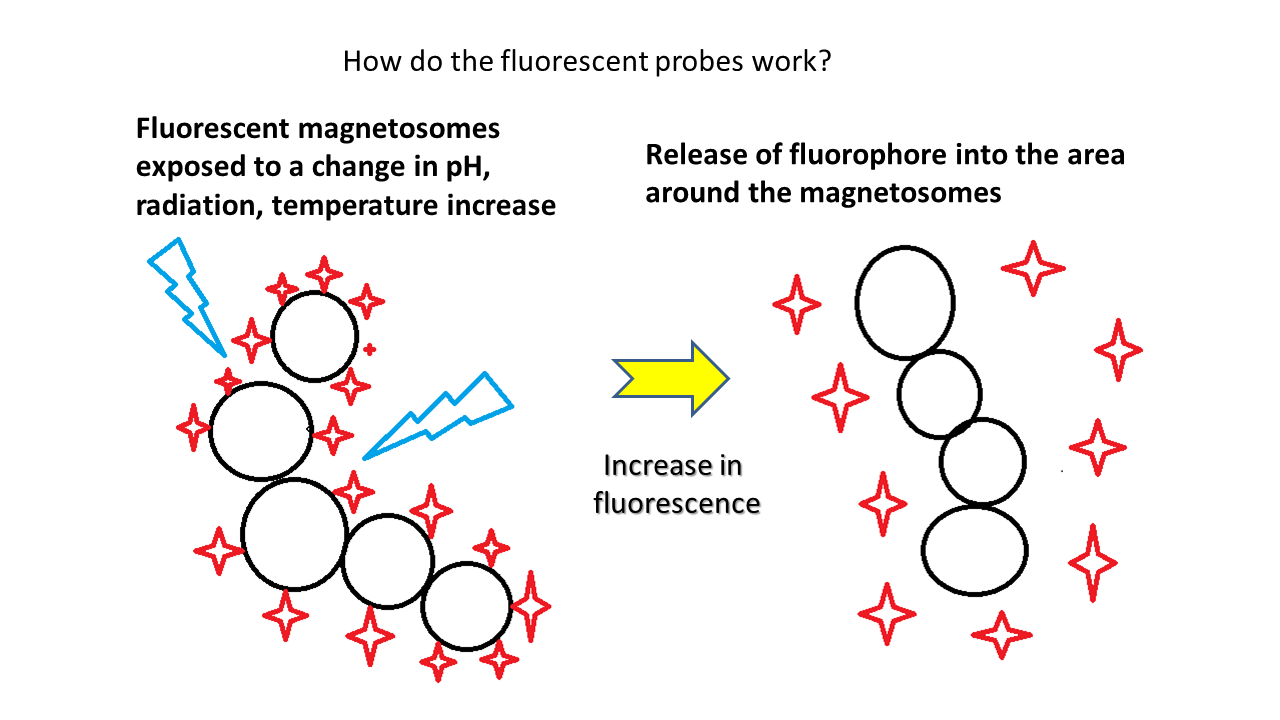
The operating mechanism of the fluorescent magnetosomes differs from that of most fluorescent probes. It relies on the dissociation of the fluorescent molecules from the magnetosomes under the application of a disturbance (radiation, change of pH or temperature). Following this dissociation, the fluorescence of rhodamine B initially attenuated by the proximity of the iron oxide increases.
The figure below summarizes the operating principle of the probe. Initially, the fluorescence of magnetosomes is attenuated. Under the effect of a disturbance (radiation, pH change, temperature increase), rhodamine B molecules dissociate from the magnetosomes to produce an increase in fluorescence.
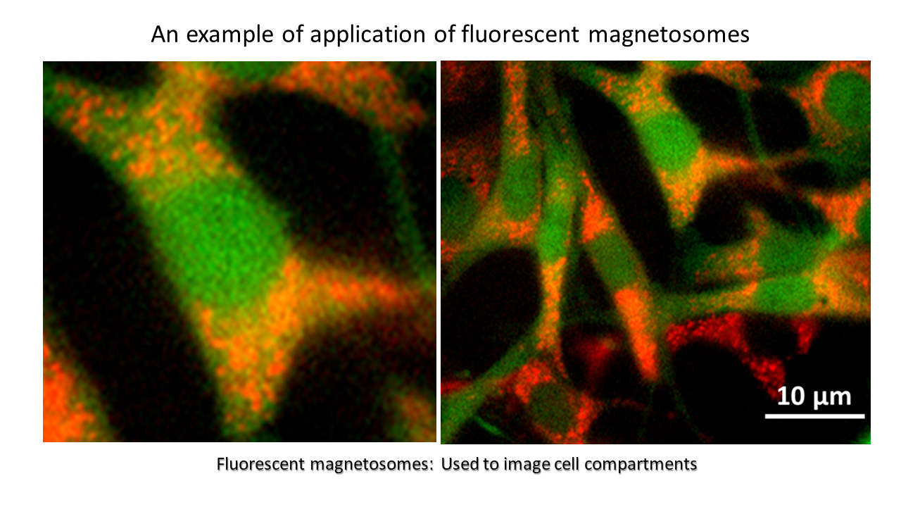
3. An example of the use of fluorescent probes: cell compartment imaging
When the fluorescent probe is added to cells and becomes internalized in the acidic cellular compartments, this induces a dissociation of rhodamine B from the magnetosomes and an increase in fluorescence.
The figure below shows fluorescent magnetosomes internalized in cell compartments with increased fluorescence intensity due to dissociation of rhodamine B.